

Our Electrophysiological Setup
- Stereotaxic apparatus for brain surgery/implantation
- Electrophysiological recording chamber
- Real-time EEG, EMG and video recording system
- Local field potential (LFP) recording system
Research Application
- In vivo brain electrophysiology
- Seizure evaluation by electroencephalogram (EEG)
- Sleep-wake polysomnographic EEG
- Electromyography (EMG)
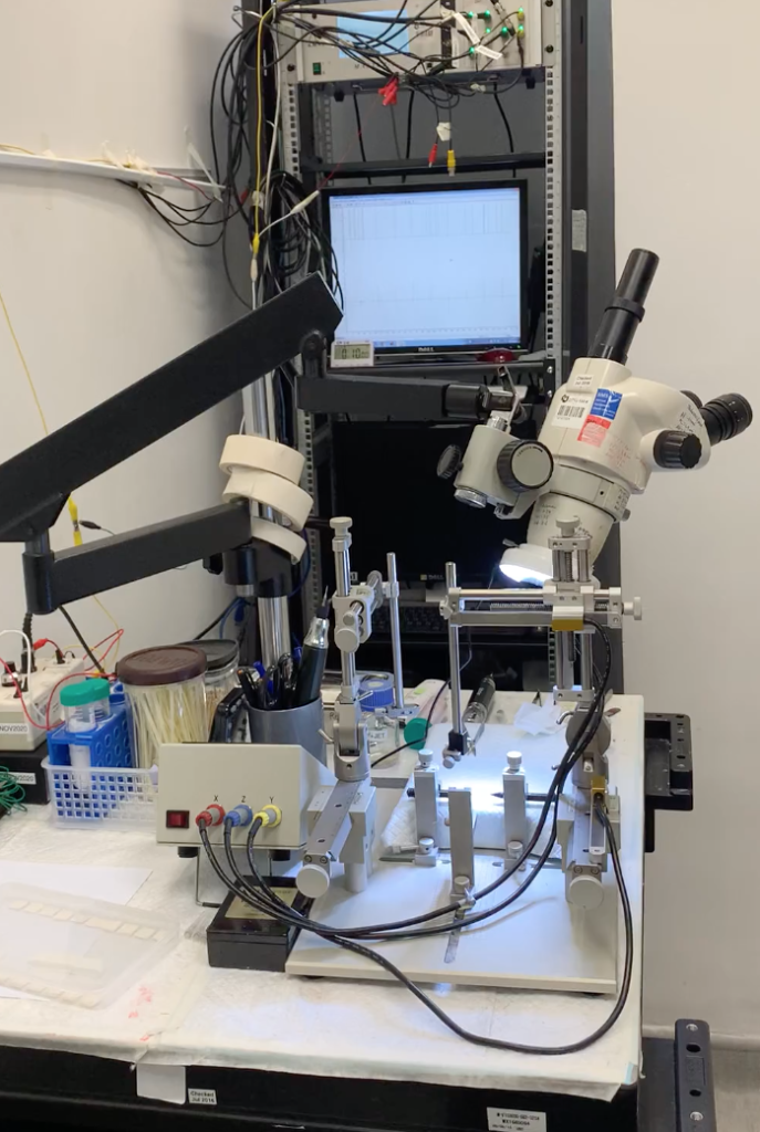
Stereotaxic apparatus for brain surgery/implantation
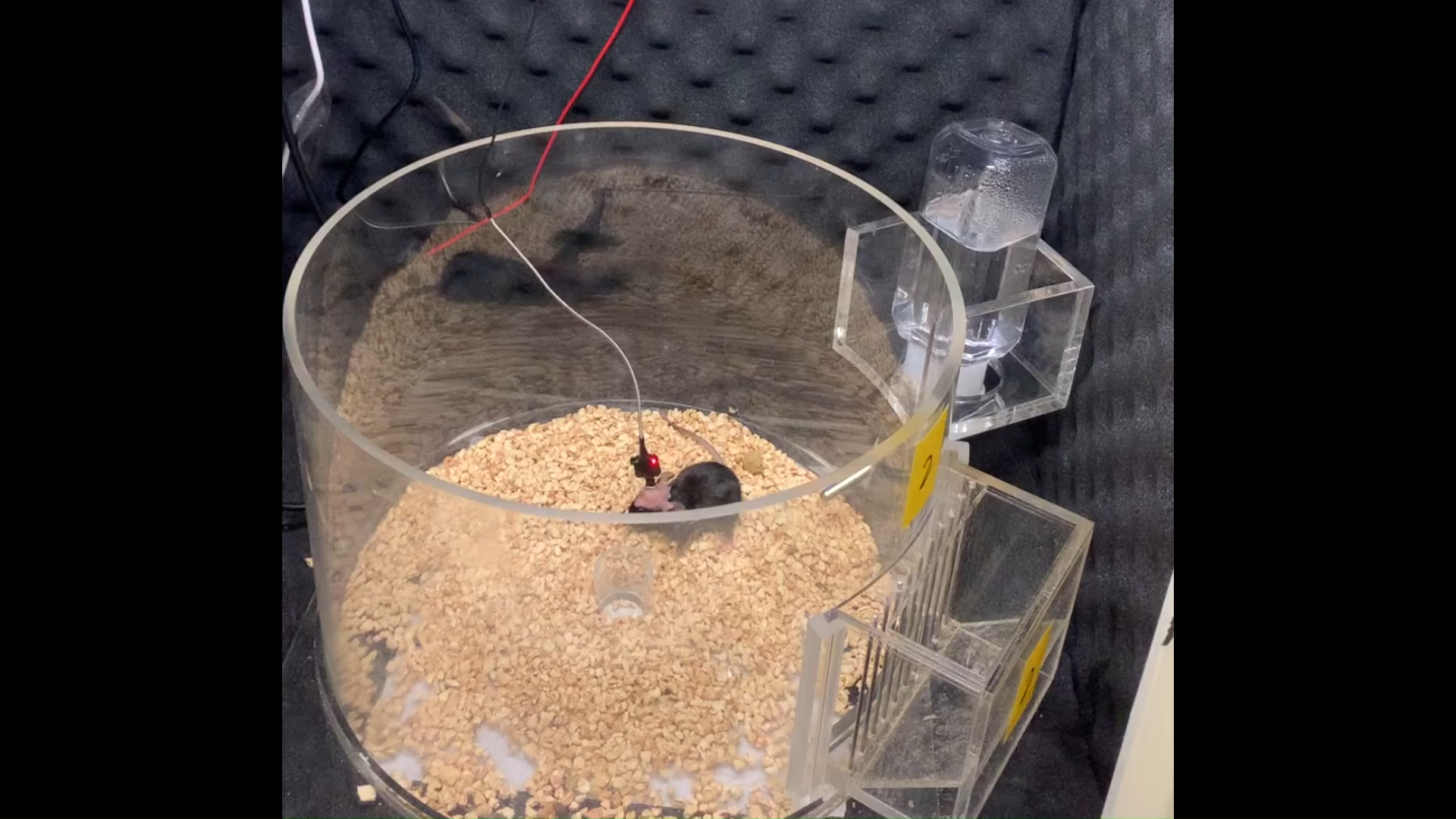
Electrophysiological Recording Chamber
The subject will be placed in a chamber with a standard light–dark cycle with food and water provided.
Real-time EEG, EMG and Video
Recording System
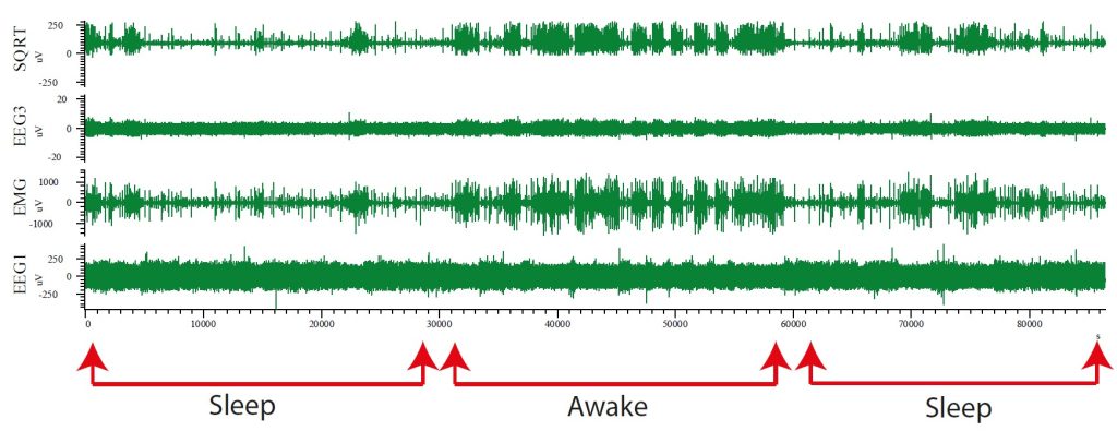
Synchronized EEG, EMG and video recordings are used to monitor the animal behaviour to evaluate the sleep stages as well as seizure severity in epileptic mice. The process will be recorded and analyzed in real-time.
The analysis can give an overview of mice’s sleep structures and sleep/wake states, as well as the seizure severity.
Local field potential (LFP) recording
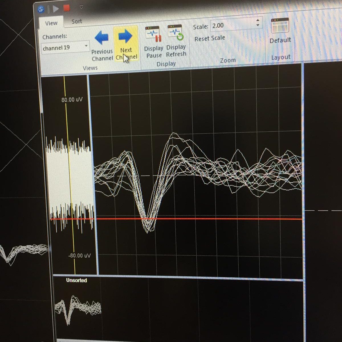
Local field potential (LFP) recordings are used to measure the neural spiking response of neurons in the subcortical brain regions of freely-moving and sedated mice.
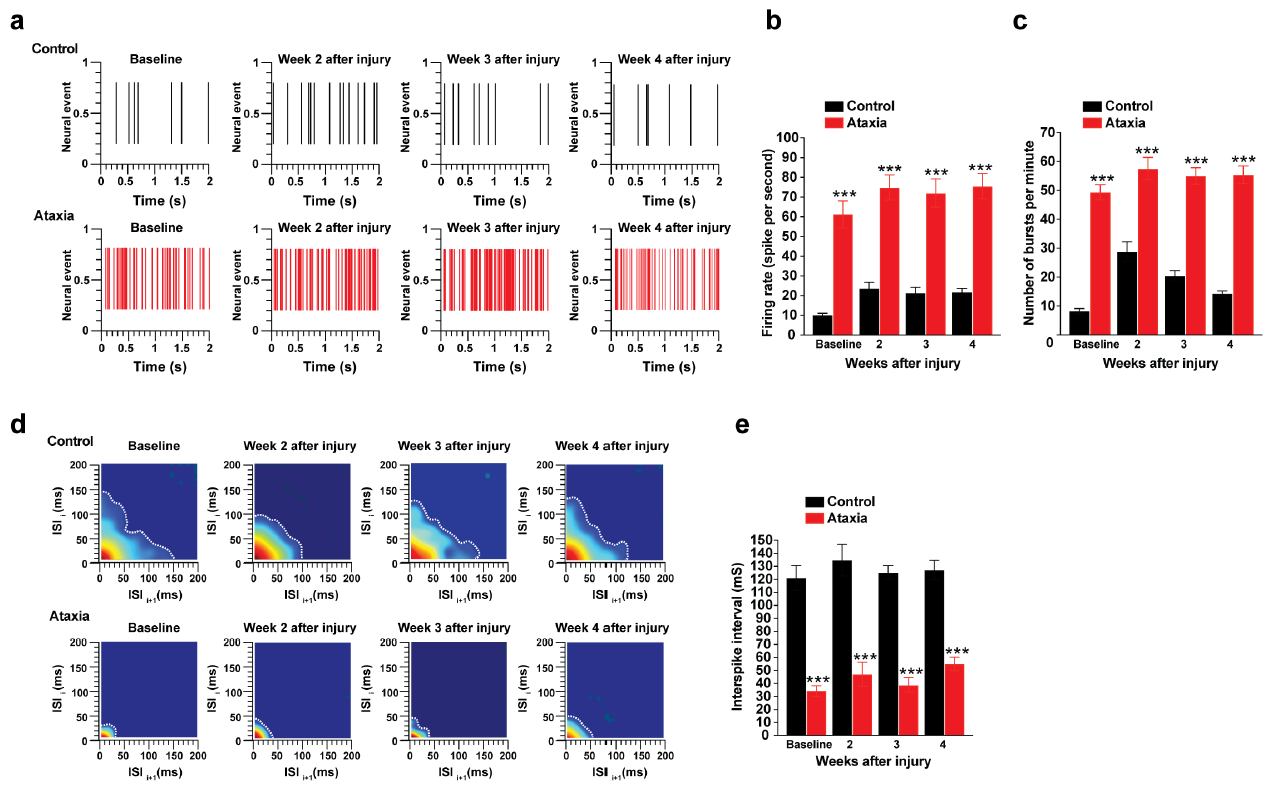
ASTHANA, P., KUMAR, G., MILANOWSKI, L. M., AU, N. P. B., CHAN, S. C., HUANG, J., FENG, H., KWAN, K. M., HE, J., CHAN, K. W. Y., WSZOLEK, Z. K. & MA, C. H. E. 2022. Cerebellar glutamatergic system impacts spontaneous motor recovery by regulating Gria1 expression. NPJ Regen Med, 7, 45.
In vivo Brain Electrophysiology
In vivo electrophysiology measures neuronal activity in the brain as either local field potentials or single units. Brain regions are targeted precisely and the effect of test compounds can be assessed. In vivo electrophysiology can be used to correlate the real-time effects of drug candidates on neuronal activity with the plasma concentration of the drug. It can also be used to detect the early biomarker of psychiatry and neurodegenerative disease such as depression and epilepsy for early diagnosis.
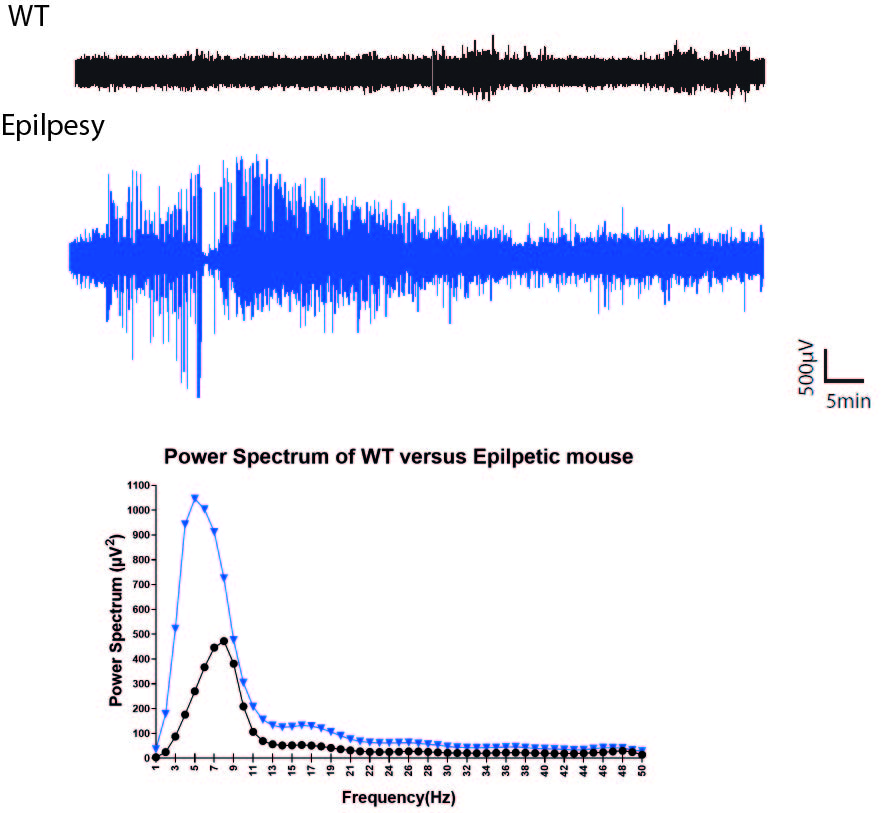
Seizure Evaluation by Electroencephalogram (EEG)
An electroencephalogram (EEG) is a neurophysiological assay used to record the electrical signals produced by the brain. Using EEG electrodes placed over the scalp surface, the electrical signal is captured and recorded.
In humans, scalp EEG is a key tool for non-invasive recording of brain activity and the diagnosis of epilepsy. EEG monitoring is also widely employed in rodent models to track epilepsy development and evaluate experimental therapies and interventions.
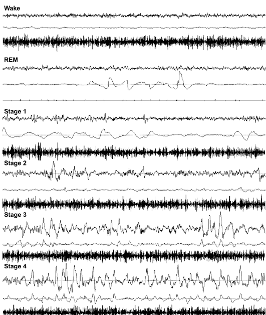
Typical human polysomnographic recordings including EEG (upper trace), EOG (middle trace) and EMG (lower trace) in each sleep-wake stage.
Sleep-Wake Polysomnographic EEG
Sleep is a fundamental biological rhythm involving the interaction of numerous brain structures and diverse neurotransmitter systems. And sleep is essential for normal cognitive function and overall health and is an important consideration in the development of novel therapeutics for many central nervous system disorders.

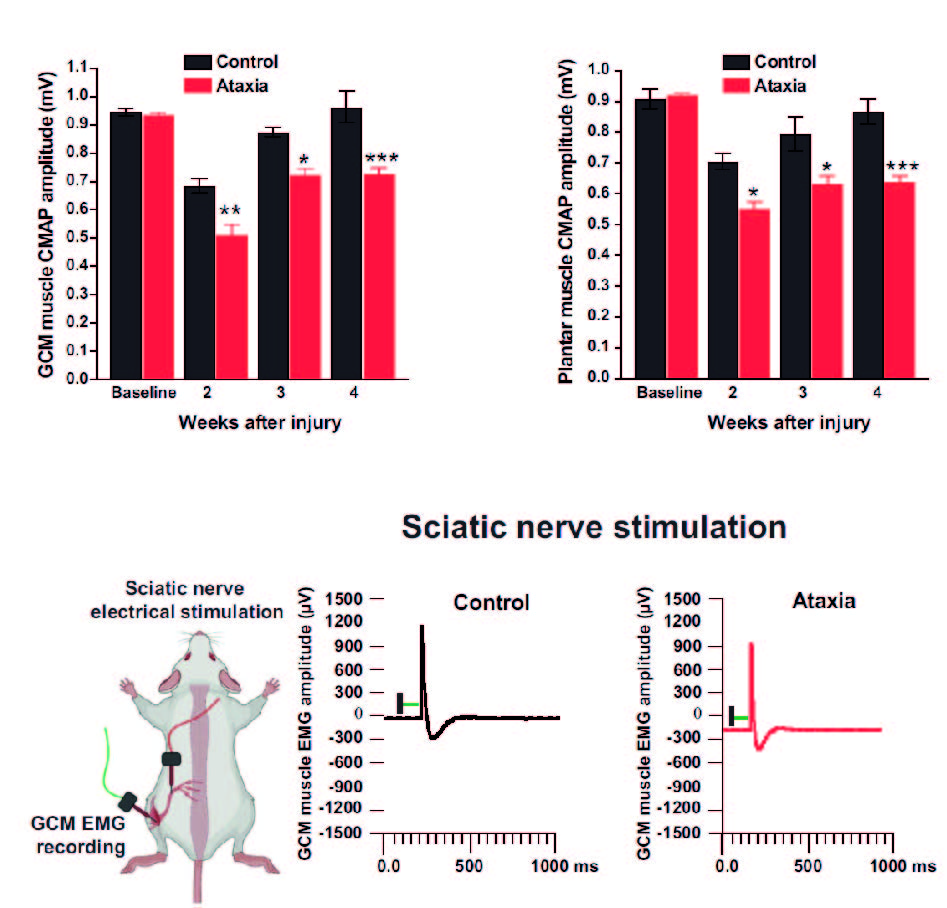
ASTHANA, P., KUMAR, G., MILANOWSKI, L. M., AU, N. P. B., CHAN, S. C., HUANG, J., FENG, H., KWAN, K. M., HE, J., CHAN, K. W. Y., WSZOLEK, Z. K. & MA, C. H. E. 2022. Cerebellar glutamatergic system impacts spontaneous motor recovery by regulating Gria1 expression. NPJ Regen Med, 7, 45.
Electromyography (EMG)
Electromyography (EMG) is a diagnostic procedure to assess the health of muscles and the nerve cells that control them (motor neurons). It measures muscle response or electrical activity in response to a nerve’s stimulation of the muscle, which can be used to detect neuromuscular abnormalities.